

Description
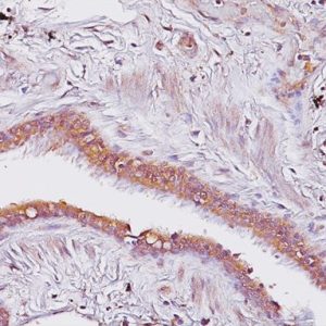
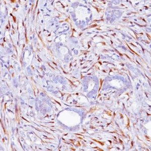
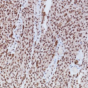
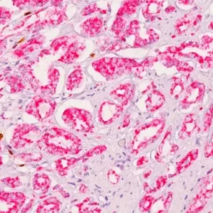
Ki-67 + Caspase-3 can provide information on cell death vs. cell proliferation in the same tissue section. The Ki-67 nuclear antigen is associated with cell proliferation and is used to grade proliferation rates of tumors. Ki-67 is found throughout the cell cycle that includes the G1, S, G2, and M phases; but not the G0 phase. Apoptosis has gained central importance in the study of many biological processes, including neoplasia, neurodegenerative diseases, and development. Cleaved caspase-3 detects endogenous levels of the large fragment of activated caspase-3, a protease that mediates apoptosis. Caspase-3 does not cross react with other cleaved caspases.
SPECIFICATIONS
Specifications
| WEIGHT | N/A |
|---|---|
| DIMENSIONS | N/A |
| INTENDED USE | IVD |
| SPECIES REACTIVITY | Human |
| SOURCE | Mouse Monoclonal, Rabbit Polyclonal |
| CLONE | DVB-2 |
| ISOTYPE | IgG1 (Ki-67 only) |
| ANTIGEN | Ki-67 and Caspase-3 |
| LOCALIZATION | Ki-67: (nuclear): brown Caspase-3: (cytoplasmic / nuclear): red |
| POSITIVE CONTROL | Tonsil or Colon Cancer |
DATASHEETS & SDS
| Download DS Data Sheet |
| Download RUO Data Sheet for International |
| Download SDS Sheet |
Regulatory Notice: Biocare’s IVD-labeled products comply with US-FDA and European IVDD regulation. Other regions may have additional requirements for such labeling, please contact your local distributor.
REFERENCES
1. Gown AM, Willingham MC. J Histochem Cytochem. 2002 Apr; 50(4):449-54.
2. Bouzubar N, et al. Br J Cancer. 1989 June; 59(6):943-7.
3. Brown RW, et al. Clin Cancer Res. 1996 Mar; 2(3):585-92.
4. Veronese SM, et al. Cancer. 1993 Jun; 71(12):3926-31.
5. Wang L, et al. Zhong Nan Da Xue Xue Bao Yi Xue Ban. 2008 Mar; 33(3):222-6.
6. Chrysomali E, et al. Oral Surg Oral Med Oral Pathol Oral Radiol Endod. 2003 Nov; 96(5):566-72.

Reviews
There are no reviews yet.